Department of Anatomy


Message from the HOD
Introduction
The Department of Anatomy at Army Medical College, Rawalpindi, established in 1977 for MBBS and later expanded in 1998 for BDS, is one of the oldest and most respected academic units of the institution. Spanning nearly 13,000 square feet of purpose built facilities, it provides a comprehensive environment for undergraduate and postgraduate education. The department trains undergraduates and post graduate students, making it a center of multidisciplinary anatomical learning.
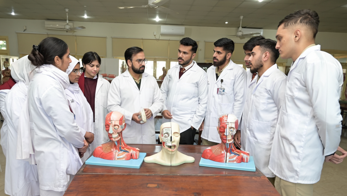
Students are actively engaged in interactive learning approaches such as focused group discussions and case-based sessions, which promote participation and critical thinking. Research activities are conducted under close academic guidance and postgraduate scholars are supported in publishing their work in well recognized national and international journals.
By combining traditional anatomical sciences with modern digital tools and plastinated teaching models, the department ensures that students gain a holistic view of the human body. Integration with Physiology and Biochemistry further enhances conceptual understanding, while clinical correlations prepare students for real world medical challenges.
Facilities & Resources
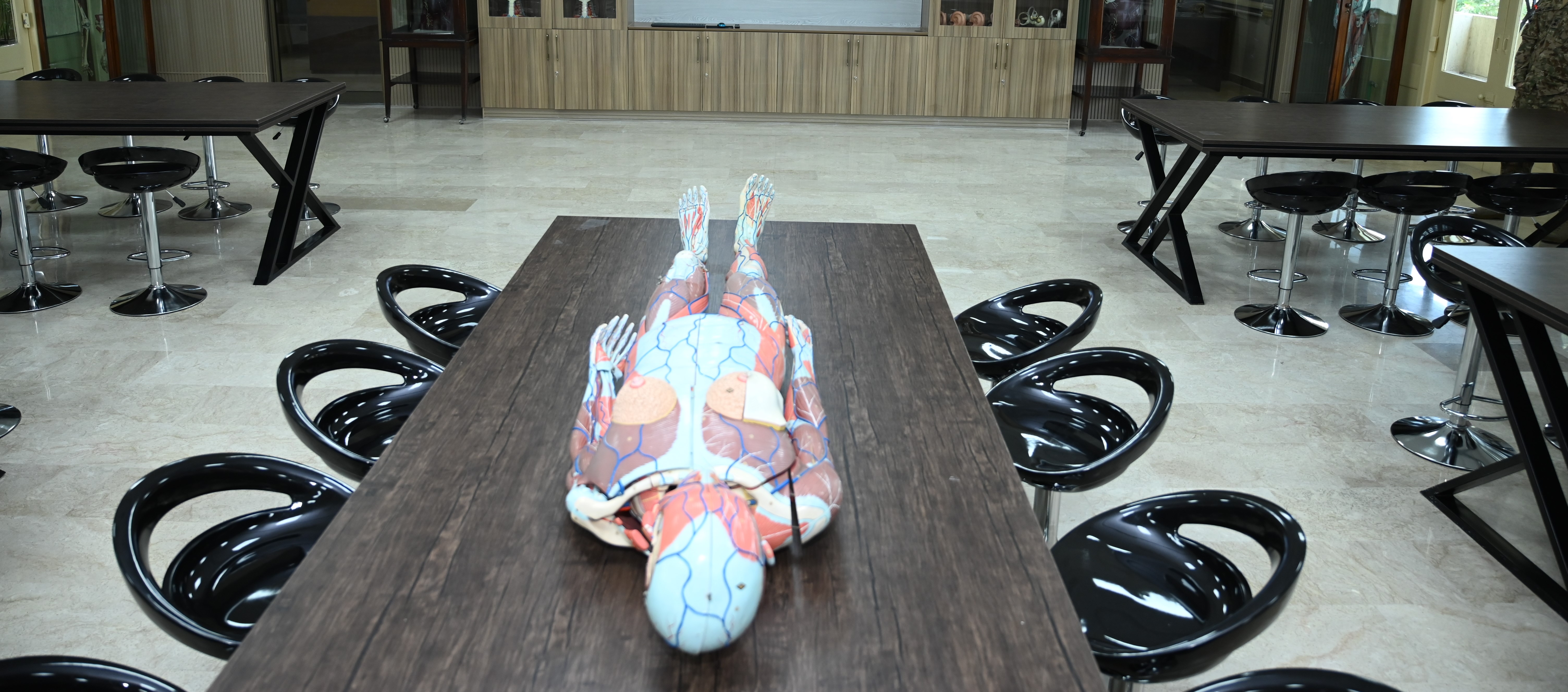
- • Plastinated Body Specimens – replacing traditional cadaveric dissection, providing permanent and detailed teaching models.
- • Histology Laboratory – equipped with high resolution microscopes and digital slide viewing systems.
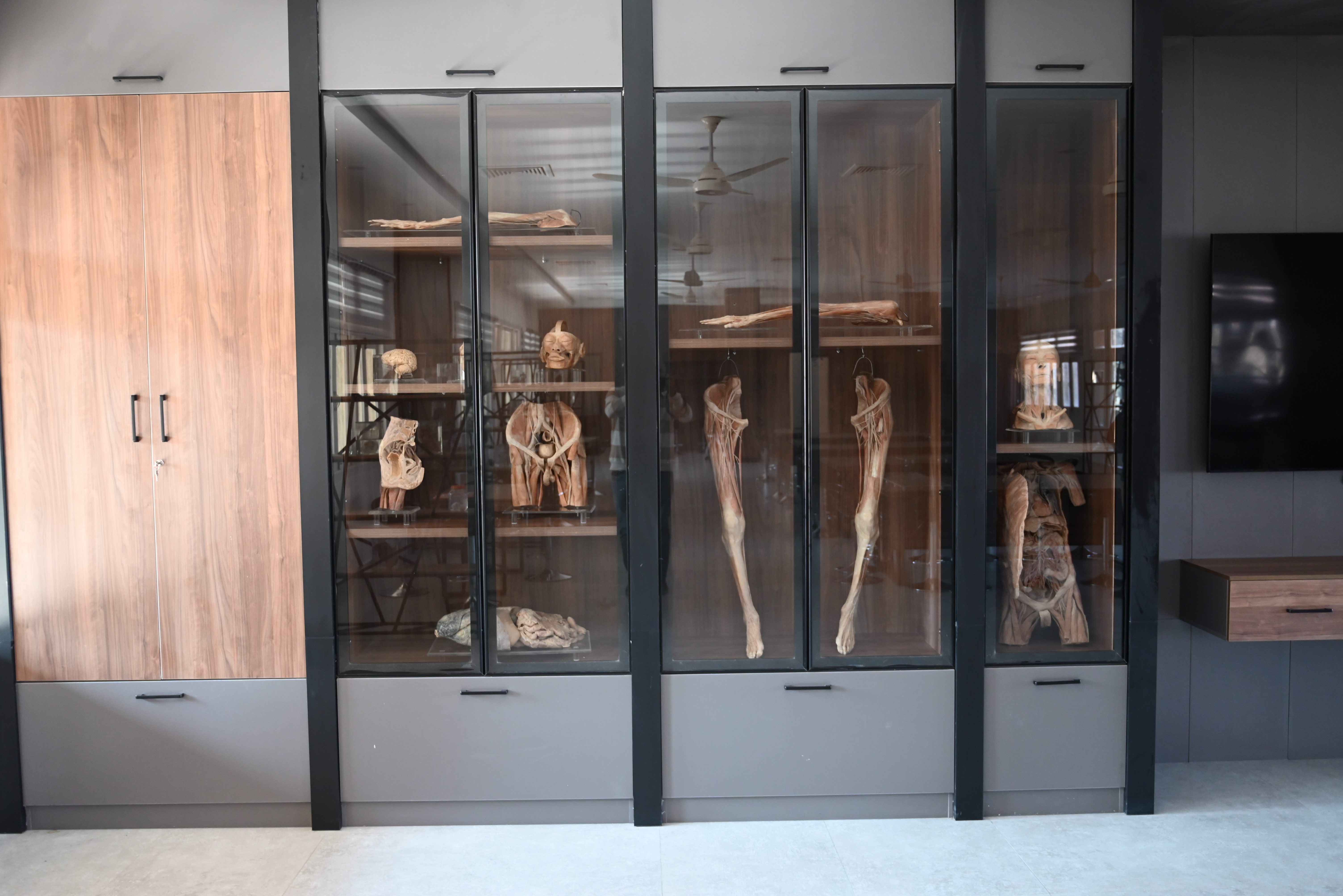
- • Anatomy Museum – curated with prosected organs, 3D Models, radiological imaging and original human bones for long lasting learning.
- • Lecture Halls – modern audio visual systems to support interactive teaching.
Outline of the Course
Anatomy at Army Medical College is taught in a systems-based, integrated format during the first two years of MBBS and first year BDS programs. The curriculum covers:
- • Gross Anatomy – Plastinated specimens, prosections, bones, models, and radiological imaging.
- • Histology (Microscopic Anatomy) – Study of cells and tissues using digital microscopy and prepared slides.
- • Embryology (Developmental Anatomy) – Human development using 3D Embryological models, congenital anomalies and their clinical relevance.
- • Neuroanatomy – Organization and function of the nervous system with applied correlations using brain specimens and models.
Teaching is delivered through lectures, practicals and interactive discussions. Case Based Learning (CBL) is emphasized to strengthen reasoning and problem-solving skills. Integration with Physiology and Biochemistry ensures a complete understanding of structure and function while correlation with clinical sciences enhances application in patient care.
Assessments include continuous evaluations, written exams, viva voce, and Objective Structured Practical Examinations (OSPE). Postgraduate students (MPhil, FCPS, PhD) receive further training in research methodology and publication ethics.
By the end of the course, graduates are expected to demonstrate not only a strong foundation in anatomical knowledge but also the ability to apply it confidently in clinical care, surgery and research, laying the basis for lifelong excellence in medicine.
Our Faculty
2
Associate Professors
4
Assistant Professors
9
Demo